All published articles of this journal are available on ScienceDirect.

Muscle Activation and Torso Movement during Exercise using Novel Fiberglass Resistance Poles
Abstract
Background:
A novel form of functional training utilizes flexible fiberglass poles for resistance. Similar to elastic bands, as the poles flex, resistance increases. To date, no studies have examined activation patterns associated with such implements.
Objective:
This study examined muscle activation and torso rotation using different pole resistance intensities during a “push-pull” rotational core exercise.
Methods:
Twenty-one subjects (16 women, 5 men; age=20.4±1.3y) completed 6 trials of 10 repetitions each of a standing push and pull movement with 3 different pole tensions (very light, light, moderate). Muscle activation (electromyography) for the anterior and posterior deltoid, abdominal oblique, and paraspinal muscles were recorded. Concentric contractions during the push phase (PUSH) and the pull load (PULL) phases were recoded, and percent maximal voluntary contraction (%MVC) was computed. Markers on the acromion process and a vertically mounted camera were used to record torso rotation during each push and pull. ANOVA for each muscle and PUSH and PULL was used for comparisons across pole intensity.
Results:
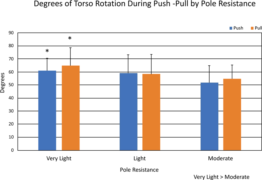
Significant main effects for torso rotation were seen, with rotation with the very light pole (Push= 61.9 ± 9.20, Pull= 64.8 ± 14.00) significantly greater than moderate (Push= 52.0 ± 12.80, Pull= 54.9 ± 10.10). EMG data were highly variable, with no differences in muscle activation detected across pole resistance loads.
Conclusion:
Variability of the EMG data prevent clear resolution of activation patterns. However, torso rotation is limited with heavier pole resistance since increased pole flex also increases resistance.
1. INTRODUCTION
Functional performance of a range of daily activity and athletic skills requires that core musculature provide a stable base during movements such as running, jumping, and sudden changes in position. Strengthened core musculature also assists with the translation of forces during sequential activity, such as translating lower body impulses to assist upper body efforts. As such, weakened core muscles can lead to falls, abnormal spinal loading during walking or running, and reduced performance of complex athletic movements [1, 2]. Training to improve functional strength requires performing exercises that overload the muscle while also allowing for various functional movements. The use of free weights or machines during exercise often prevents variations in angle, speed, and torso movements necessary for functional training.
Alternative forms of resistance training have been developed using elastic bands or flexible materials, which overcome these limitations [3-5]. Andersen et al. [5] found similar loading was possible between elastic bands and dumbbell exercises, with intensities between 60-80% MVC. Resistance bands offer unique options for functional rehabilitation due to their ability to be used in a variety of movement planes and still effectively strengthen the muscle [6-9]. The bands are color-coded, with each color representing different resistance properties, creating a range of loading schemes. The amount of resistance elastic materials provide is dependent on the stretch deformation characteristics. Generally, the amount of tension provided increases proportionally with the amount of deformation of the band. Hughes et al. [10] determined the resistance properties of 6 progressive levels of Thera-Band™ tubes during shoulder abduction exercise. The study showed a positive, linear relationship between tube length and tension, with significant tension differences between the colored tubes. Uchida et al. [11] published data showing tension for Thera-Band™ elastic bands at strain values between 25% and 250% of resting length. By quantifying the resistance of these bands, these data provided useful information to practitioners that allow them to appropriately prescribe loads for training.
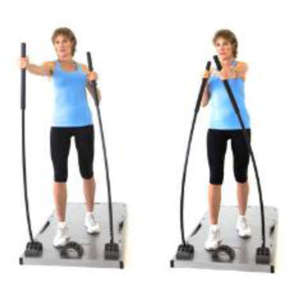
More recently, resistance pole technology has been developed that also provides resistance through a range of planes. Core Stix™ is a relatively new resistance training technology [12] currently available and marketed as a training technology for both functional sports training as well as rehabilitation. The training system consists of a platform with metal interfaces that contain sockets on the left and right sides of the platform-oriented in a variety of directions (Fig. 1). Fiberglass polymer resistance poles can be inserted into the sockets and flexed through a range of motion, creating tension as they are flexed. As the poles flex, there is a linear increase in resistance. Glass and Remski [13] have quantified the load provided across the flex range of the poles, with ranges from 0.8kg to 40kg depending on the flexion, type of pole, and handhold [13]. Five different pole intensities exist, differentiated by colors. Intensities range from purple (very light), white (light), yellow (moderate), blue (heavy), to red (very heavy). The handhold area of the poles is large (44cm), so variations in hand placement do influence the amount of tension within the same pole, such that the bottom of the handhold area provides 1.7 times the resistance of the top handhold area. To date, there is no published research regarding this new technology relative to the efficacy of activating core muscles during a functional movement. Since functional movements require a range of motion that may potentially be impeded by a device that increases resistance as the movement continues, it is important to understand how different loading schemes affect a movement designed to activate core muscles. The purpose of this study was to utilize a common push-pull action using three different pole intensities (“very light, light, moderate”) and assess the activation of limb and core musculature as well as a range of motion.
2. MATERIALS AND METHODS
2.1. Subjects
For this study, we recruited healthy, active men and women over the age of 18 from a University population. Twenty subjects completed all testing (15 women, 5 men; Age = 20.4 ± 1.3y, Height = 170.2 ± 9.8 cm, Mass = 66.1 ± 13.6kg, systolic blood pressure = 118.7 ± 12.5 mmHg, diastolic blood pressure = 75.5 ± 10.8 mmHg). All subjects provided written, informed consent, and the study was reviewed and approved by the Institutional Review Board for the University.
2.2. Initial Screening, Orientation, and Practice
After the protocol was explained and subjects provided written consent, subjects completed a health history questionnaire [14], and resting blood pressure was measured. Inclusion criteria consisted of indicating moderate physical activity at least 3 times per week, no orthopedic limitations and resting blood pressure under 130/80 mmHg. Height (SECA 222 wall-mounted stadiometer, Chino, CA) weight (SECA 500 beam scale, Chino, CA) and manual blood pressure were measured prior to orientation.
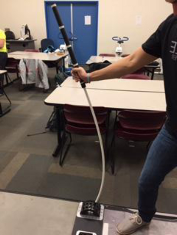
The orientation session introduced subjects to the Core Stix apparatus. Subjects watched a brief video provided by the manufacturer [12] showing rod placement, proper foot placement and technique for the “Push-Pull” exercise (Fig. 1). Due to the expanded size of the hand hold area, 4 sections were marked on the handles, and subjects practiced with hand placement until finding a hold that felt most comfortable for them based on their body size. Due to variations in resistance due to hand hold placement [13], subjects were required to maintain the same handhold setting (Fig. 2) throughout the study. Subjects practiced the push-pull movement using all three resistance poles (Purple- very light, white- light, yellow- moderate), executing the push-pull action. Cadence was controlled using a metronome and was set at 18 repetitions per minute. Subjects practiced in 15s increments.
2.3. Experimental Treatment
Within 48 hours of the orientation session, subjects reported to the lab for the experimental session. Subjects completed a total of 6, 30s exercise trials across the 3 resistance poles. The order of treatment was counterbalanced. Electrodes were placed on the right side of the body, and due to the push pull aspect of the exercise, two trial per pole were completed, and the positions of the poles were reversed, allowing a “push-pull”, and a “pull-push” movement to be assessed. In order to assess trunk rotation, the overhead video was recorded during the trials.
2.3.1. Electrode Placement
Bipolar, pre-amplified, active surface electrodes (Biopac TSD150B, Biopac Systems Inc., Goleta, CA) were placed on the right side of the body according to locations identified by Cram [15] following shaving and cleaning with alcohol. Muscles assessed included: anterior deltoid, posterior deltoid, abdominal oblique and paraspinal. A single ground electrode was placed on the sternum and connected to the Biopac MP 150 system. Data were collected at 2,000 samples per second.
2.3.2. Torso Rotation
To assess the degree of torso rotation as a result of the push-pull motion, reflective markers were placed on the left and right acromion processes of each subject. A video camera was placed overhead at the height of 2.44 m of the subject so that each repetition could be recorded for later measurement. The view provided a 90-degree viewpoint, making an accurate 2D assessment of rotation possible (Fig. 3).
2.3.3. Exercise Trials
Treatments were applied in counterbalanced order for each subject. Subjects completed 8-10 submaximal repetitions across 30s for each pole intensity, with poles reversed to sample both push and pull for the right side of the body. This required a reversal of foot placement as well as starting position (either from a push or pull position). A minimum of 3 minutes of rest was allowed between each set. Cadence was maintained by a metronome as close as possible by the subjects. Due to the variable movement of the poles during the activity, which precluded the use of an electronic switch, each repetition was marked visually by a researcher during the trials on the EMG recording system, allowing assessment of both concentric and eccentric contractions. Handhold setting varied across subjects but was held constant for each individual subject according to their selection. Following exercise trials, each muscle was assessed individually for maximal voluntary contraction (MVC), while EMG was recorded. Isometric maximal contractions against a brace were performed using shoulder flexion (anterior deltoid), shoulder extension (posterior deltoid), angled curl up (abdominal oblique) and reversed curl-back extension (paraspinal).
2.4. Data Reduction
EMG data were filtered using a high pass filter (Blackman – 67 db), rectified, and integrated (IEMG). Due to the variability of movement speed, IEMG values were corrected to account for differences in sample size. Final IEMG data were converted to percent MVC based on IEMG from the MVC tests. For the purposes of the present study, only concentric data were assessed.
Torso rotation images were viewed and assessed for degrees of rotation using KINOVEA open-source movement analysis software [16]. Data for each repetition of the push and pull motions were measured for each pole resistance. The subject’s starting point in the push pull position was used as the initial position, and a line between acromial markers used as the starting zero point. Change in position relative to the left acromial point was measured in degrees by advancing frame by frame to identify the end of each movement using a standard laptop computer (Lenovo Thinkpad T450, Hong Kong, China) and wireless mouse. Software tools allowed a measurement of the rotating relative to the acromial marker. Past research on the validity of the KINOVEA software suggested that a 90 degree angle for camera position provides the most valid measure of 2-dimensional movement such as rotation [17].
Statistical analysis was assessed for torso rotation using a one-way analysis of variance for push and pull movement separately (muscle, pole resistance, muscle x pole resistance). When significant main effects existed, post hoc Tukey t-tests were used for comparisons. EMG data were assessed within each muscle group across pole intensities also using a one-way ANOVA.
3. RESULTS
Similar to exercise bands, fiberglass resistance poles increase resistance as they are flexed, creating conditions of constant adjustment to added loads as the individual progresses through a range of motion. Fig. (3) shows the degree of torso rotation across the resistance pole intensities. Significant differences existed in the degree of torso rotation, with subjects performing significantly greater torso rotation during both the push and pull movement for the very light pole resistance (Push = 61.1 ± 9.20; Pull = 64.8 ± 13.60) compared to the moderate (Push= 52.0 ± 12.8; Pull = 54.9 ± 10.10).
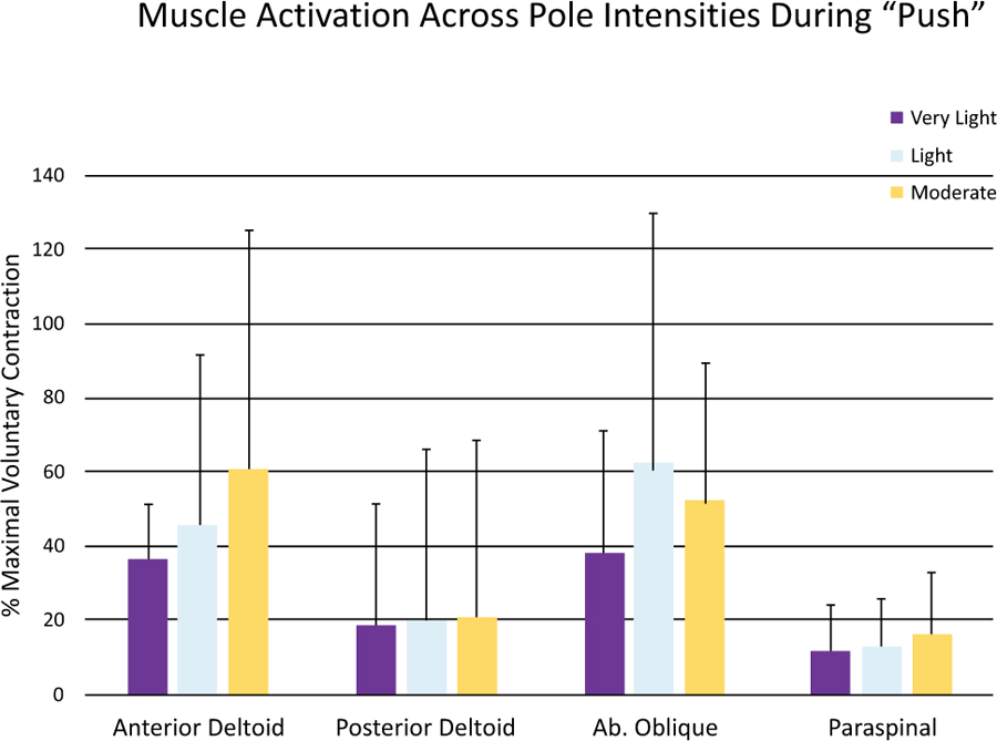
Due to very wide ranges in EMG activity, no statistical differences were seen across the push conditions for any muscle (Fig. 4 and Table 1). There were mean data trends towards more anterior deltoid and abdominal oblique activation as the pole resistance increased, with very wide standard deviations.
| - | Push | Pull | ||||
|---|---|---|---|---|---|---|
| Muscle | Very Very Light | Light | Moderate | Very Very Light | Light | Moderate |
| Anterior Deltoid | 36.67 ± 14.67 | 45.90 ± 45.87 | 61.10 ± 64.03 | 33.54 ± 28.29 | 32.22 ± 34.36 | 29.30 ± 28.89 |
| Posterior Deltoid | 18.80 ± 32.63 | 20.09 ± 45.96 | 20.46 ± 47.98 | 40.73 ± 102.21 | 25.90 ± 58.21 | 31.19 ± 53.20 |
| Abdominal Oblique | 38.01 ± 32.73 | 63.24 ± 66.09 | 52.89 ± 36.19 | 32.19 ± 22.71 | 52.35 ± 51.58 | 49.03 ± 29.36 |
| Paraspinal | 11.90 ± 12.34 | 13.08 ± 12.73 | 15.96 ± 16.94 | 22.30 ± 16.92 | 24.78 ± 18.18 | 35.03 ± 30.76 |
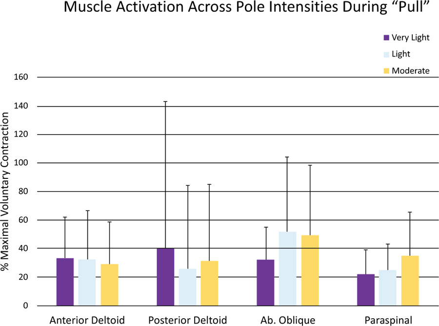
Pull data were also highly variable, making statistical analysis difficult (Fig. 5). Mean data (Table 1) suggested that posterior deltoid activation during the pull was favored more with the very light pole, and at the moderate pole resistance deltoid activation was reduced as paraspinal activity increased. One might speculate that the increased pole resistance precludes the involvement of the small posterior deltoid and emphasizes larger back muscle activation. This may explain why the range of motion was also reduced.
4. DISCUSSION
Functional training is widely used in both sport and therapeutic applications in order to provide a variety of movement possibilities and lead to improvements in functional performance and core muscle stability [17]. A variety of modalities have been employed, such as suspension exercises [18-20], instability training [18], and elastic bands and tubes [5, 8, 21]. The present study is unique in that it is the first to examine a new modality of functional training utilizing flexible fiberglass poles [12]. Our results indicate that the fiberglass poles provide a substantial resistive load for the limbs and torso during a “push-pull” exercise using three different pole intensities. As the load intensity increased, the range of torso rotation was significantly reduced due to the added resistance on the poles as they flexed. The resulting change in muscle activation corresponding to this reduced rotation was difficult to determine. Data varied widely, and most likely due to the different resistance relative to the strength of each individual subject. While each subject did serve as their own control, the muscle activation pattern across the three resistance intensities likely varied to the extent that some utilized greater core muscle activation and others more limb (shoulder) activation. Four different handhold options were presented to each subject, and most selected a hold based on height. However, this creates loading differences that may not be ideal for subjects by size, as often the smaller female subjects chose lower handholds, and this resulted in a larger starting load due to the higher resistance at lower hand holds [13].As a combined sample, the results were therefore varied to the point of only suggestive trends.
Rotation of the torso and pelvis, along with shoulder flexion and extension, provides three different locations for both stabilization and power generation. Vera-Garcia et al. [22] studied muscle activation patterns during isolated rotations at the pelvis or torso in trained dancers during upright standing. They found differences in muscle activation when a rotational movement was driven by the pelvis compared to the torso. Internal oblique activation was significantly higher during pelvic-driven motion, while abdominal obliques were activated to a greater extent during torso rotation. However, they noted considerable inter-individual differences, despite testing a group of trained belly dancers with the exceptional torso and pelvic control. Similar to the stretch of elastic bands, fiberglass poles increase resistance as they flex. This means that for the same pole resistance, relative resistance is continually increasing as the poles flex, and in the case of this study, as push-pull torso rotation continues. Therefore, the motion will be limited by relative strength, as well as the strength of individual muscles contributing to the movement. A weak or deconditioned muscle in the chain of movement may result in a different strategy for execution of the push-pull, indicated by the high variability in activation see in the present study. Lett and McGill [23] examined pushing and pulling technique between firefighters (experienced push and pull subjects) compared to unpracticed subjects. Subjects executed two-handed push and pull maneuvers with different loads while walking 3 steps. Results showed greater spinal loading and EMG activity in novice subjects compared to experienced firefighters. Clearly, technique influenced EMG activity, with the experts utilizing less spinal flexion and more torso flexion. In the present study, the unique nature of the fiberglass poles results in added load at the push/pull continues, creating a variety of possible scenarios for muscle activation in order to complete the movement. For example, in the present study mean data (Table 1) suggested that posterior deltoid activation during the pull was favored more with the very light pole, and at the moderate pole resistance, deltoid activation was reduced as paraspinal activity increased. With reduced torso rotation at the higher loads, it may be that pole resistance precludes involvement of the small posterior deltoid and emphasizes latissimus dorsi muscle activation. Additionally, research by Botton et al. [24] found that posterior deltoid was activated more readily during horizontal shoulder extension rather than the sagittal plane extension of the present study. With a smaller range of motion at the higher resistance, the movement becomes more restricted to the sagittal plane, with less horizontal extension, so this may limit posterior deltoid action. Similar results were seen by Bennett et al. [25] with single and two-handed push and pull movements. EMG activity varied considerably due to individual differences in technique. Since the poles allow varied movement through different planes, activation may change to favor the largest muscles.
While hand setting was held constant, the range of motion varied among participants since relative strength varied in comparison to each pole intensity. At the lightest resistance, most individuals were able to execute a full torso rotation and shoulder flexion-extension. However as the pole resistance increased, different strategies were used to execute the push pull action. This could have caused a shifting of muscle activation, as torso rotation was reduced and braced for stability, while shoulder activation and perhaps elbow flexion-extension musculature was increased. Abdominal activation may have assisted with torso rotation at low resistance yet served more as a brace at higher resistance. The data also suggest that a lighter pole resistance, and one that allows a full range of motion, may be desirable for activation of a wider range of core muscles since added load does not appear to cause additional activation specific to core muscles. Vera-Garcia et al. [22] showed that the latissimus dorsi muscle becomes significantly more important to the rotation of the torso, however, we did not evaluate this muscle. Due to the limited number of muscles studied, it was not possible to determine if the force needed to complete the push-pull action was redistributed across a wider range of muscles as the resistance increased. It is likely that an “off-loading” of work was distributed to different muscles during the rotation movement, meaning that a push-pull motion is a far more complex activation pattern depending on the forces involved. Data showed significant reductions in rotation at the heaviest resistance, and this may explain the lack of change in abdominal muscle activation across the resistance intensities. Berquist et al. [21] studied the pectoral fly motion using free weights compared to elastic bands. Interestingly, free weights induced higher activation in the pectoral muscle, but the elastic bands elicited higher activation in the ancillary support musculature. It may be that the instability induced by the elastic flex nature of the Core Stix poles causes changes in the activation of ancillary support muscles as part of the load distribution. This is difficult to measure without assessing a much larger array of muscles.
The present study has limitations that make it difficult to fully understand the nature of muscle activation patterns across the increasing pole resistance intensities. As previously stated, the selected handhold position influenced the starting load, and smaller individuals selected a lower handhold, thereby starting with a higher load than taller subjects. While mean percent MVC values for subjects only ranged from 11% to 53% across muscles for the three poles, individual differences resulted in wide variability. Future studies with this device will need to control variables such as height, handhold and initial strength along with a wider array of muscles investigated to follow the changes in recruitment patterns as the loads change. It is clear, however from the present study that lower resistance results in a complete push-pull movement and more predictable muscle recruitment. As resistance increased and different strategies were employed to complete the movement, there was not a consistent change in recruitment pattern. To safely recruit core musculature and execute the push pull movement, it is recommended that light resistance be used for the safest movement.
CONCLUSION
Stabilization and movement of the core during functional exercises is a complex and changing pattern. Our data show that as pole resistance is increased to the degree of torso rotation declines. As a result of this decline, there may be various individualized strategies used by subjects in order to execute a movement. This may mean core muscles that had been used to drive torso rotation at a lower resistance were instead used as stabilizers at a higher resistance, while limb muscles were used more to complete the movement. Results suggest that for a full range of motion utilizing core muscles, one should maintain a low enough resistance to execute a full range of motion.
ETHICS APPROVAL AND CONSENT TO PARTICIPATE
This study was reviewed and approved by the Grand Valley State University (MI, USA) Institutional Review Board for human subjects research (# 17-011-H).
HUMAN AND ANIMAL RIGHTS
No animals were used in this research. All human research procedures followed were in accordance with the ethical standards of the committee responsible for human experimentation (institutional and national), and with the Helsinki Declaration of 1975, as revised in 2013.
CONSENT FOR PUBLICATION
All participants signed informed consent prior to participating in the study.
STANDARDS OF REPORTING
STROBE guidelines and methodologies were followed in this study.
AVAILABILITY OF DATA AND MATERIALS
De-identified subject data are maintained by institutional archives.
FUNDING
None.
CONFLICT OF INTEREST
The researchers declare no conflicts of interest with this study, financial or otherwise.
ACKNOWLEDGEMENTS
I would like to acknowledge the student research assistants who helped throughout this research study. Particular acknowledgement to Mr. Steven Zanders, Ms. Jessica Montalbano, and Mr. Jared Gordon.