All published articles of this journal are available on ScienceDirect.

Relationship Between Knee Extensors Power Output and Vastus Lateralis EMG Activation in Elderly Women: Influence of Mother Wavelet Selection
Authors Info & Affiliations
Abstract
Purpose:
The main objective of this study was to compare frequency parameters produced by six mother wavelets pinpointing the most feasible to investigate electromyographic (EMG) parameters while producing knee extension power in elderly women. The influence of different load conditions in mother wavelet selection and power output were also analyzed.
Methods:
Thirteen sedentary elderly women (69.3 ± 4.1 years) took part in the study. Participants executed 6 repetitions of 3 load condition (30%, 50% and 70% of the maximal) with the concentric phase of the knee extension movement as quickly as possible. Kinematic data obtained by video analysis, an anthropometric model and Newtonian mechanics were used to calculate knee extensors’ power. A continuous wavelet analysis was used as a time-frequency transformation strategy of vastus lateralis and biceps femoris EMG data and six different mother wavelets were selected: Morlet; 4th, 8th and 44th order Daubechie, 4th order Coiflet and 5th order Symlet.
Results:
44th order Daubechie showed the highest maximal cross correlation value and no differences were seen between different mother wavelets and cross correlation at zero lag and in the lag variable. Although increased knee extensors peak power at higher loads were seen, no differences in vastus lateralis or biceps femoris root mean square values were obtained.
Conclusion:
44th order Daubechie mother wavelet was pinpointed as the most suitable to obtain EMG time-frequency parameters. We have also seen that different load conditions do not seem to have an influence on mother wavelet selection.
1. INTRODUCTION
Muscle strength decrease as a sarcopenia consequence is strongly related with a prolonged healthcare assistance need and a reduced quality of life [1]. This physiologic section area reduction is primarily due to the diminishing muscle fibers number as well as to their size, particularly the fast twitch muscle fibers [2]. These changes seem to have severe qualitative consequences in this tissue functional performance. Greenlund and Nair [3] showed that between the third and the eighth decades the knee extensors’ ability to generate peak torque suffers a significant decline indicating an inverse relationship between aging and muscle function. This particular characteristic has been related to decreased mobility [4] as well as identified as a risk factor for falls [5].
Indeed, all of these quantitative and qualitative changes in a senescent musculoskeletal system have even more profound functional outcomes that lead to a condition in which the daily tasks once effortless performed now seem to be challenging ones [6]. Being the majority of these tasks of short duration and, because of that, intimately related with the capacity to produce force as quickly as possible it would be expected that muscle power plays a key role in older adult’s functional capacity. In fact, Brunner et al. [2] refer that among the main qualitative muscle changes, muscle power loss is the one that early occurs suggesting it as the most striking muscle alteration in the senescent locomotor system. The authors suggest that this gradual inability to rapidly produce muscle force appears to be a more markedly characteristic in women as they have a priori less muscle mass. The functional capacity diminution multifactorial origin makes the ideal exercise protocol to increase this ability to perform everyday tasks unclear. Tschopp et al. [7] pinpoint muscle power as the main independence determinant in the elderly. The authors state that when compared with muscle maximum strength, this parameter presents a premature and abrupt decline making power training not only promising but unavoidable in a senescent life. They define this method as a moderate strength training in which the concentric phase of the exercises are executed as quickly as possible and the eccentric phase in a moderate velocity. When compared with strength training, power training has been found to be superior [7-9]. Indeed, this method allows enhancing older adult’s functional capacity, as well as the conventional one, with significantly less training loads, reducing the training perceived effort.
Trying to determine the training load that allows producing the highest power output Zbinden-Foncea et al. [10] found a U-shaped trend with maximal power for loads of approximately 60% of the maximal effort (1RM), both for upper and lower limbs in elderly women. However, Signorile et al. [11] stated that optimal loads are affected by the architecture of the joints being trained. They showed that joints associated with longer bones are prone to higher training speed than those associated with shorter ones. Nevertheless, until now there is no consensus regarding the optimal load condition to produce maximal peak power and the neuromuscular factors affecting knee extensors power production in older adults seems to have not been fully clarified yet.
Merletti et al. [12] indicated that the selective atrophy of type II muscle fibers diminish the contractile properties lowering motor unit firing rates of the older muscle. The authors suggested that these are determinant factors of the maximal voluntary contraction torque as well as of the myoelectric manifestations of muscle fatigue. Indeed, when compared to older people, young people seem to have a greater rate of decrease of the power spectrum parameters in fatigue-type conditions [12, 13]. This supports the theory of elderly muscles shifting toward slow-twitch muscle fibers [14]. Besides determining muscle fatigue, frequency spectrum analysis of the EMG signal has been related, with some controversy [15, 16], to muscle fiber type [17].
Although Fast Fourier Transformation (FFT) is the most frequently used method to assess EMG frequency spectrum it is limited to a global frequency analysis, with no time resolution [18]. A time-frequency resolution of the EMG signal could provide greater insight into the relationship between muscle power output and muscle frequency rate. Wavelet Transform is a powerful technique that allows extracting EMG frequency features since is consistent with the non-stationary nature of the EMG signal, providing an alternative to the classical Short-Time Fourier Transform (STFT). While the latter strategy uses a single analysis window, the Wavelet Transform uses short windows at high frequencies and long windows at lower frequencies which allows to shift as well as distend and contract (scaling) a prototype function (mother-wavelet) without losing time resolution [19]. The selection of the proper mother wavelet applied to the biological signal to be analyzed imposes another challenge in order to understand the relationships between muscle frequency and mechanical information such as power output. Therefore, the main objective of this study was to compare frequency parameters produced by six mother wavelets pinpointing the most feasible to investigate quadriceps femoris EMG parameters while producing knee extension power. A second goal was to analyze the effect of load magnitude in the selection of the optimal mother wavelet and in knee extensors power output.
2. MATERIALS AND METHODS
Took part of the study 13 sedentary elderly women (69.3 ± 4.1 years) with similar body mass index (26.1 ± 2.5 kg/m2). They had not started any physical activity program for at least 2 years and reported absence of cardiac and musculoskeletal problems as well as arterial hypertension. The participants were informed of all the operational procedures giving their Written Consent informing that their involvement in the study was voluntary. The local Ethics Committee approved all procedures (protocol number 2010/16). Baecke questionnaire [20] modified and validated for older adults [21] was used to assess their physical activity level.
Instruments: A customized knee extensors machine was used for the experiment which allowed placing an electrode in the participants’ hamstrings without contacting the seat. A digital video camera (Casio EX-ZR10) with a sample rate of 240Hz and a shutter speed of 1/2000s recorded the trials. Reflexive markers (20 millimeters diameter) were attached to the participants’ lateral malleolus of the ankle and lateral condyle of the knee. Such markers allowed assessing knee angle in relation to the machine’s initial position. These data were used to calculate knee concentric angular velocity and acceleration. An external trigger allowed synchronize the kinematic and electromyographic data. Lynx-EMG System 1000 (Lynx Electronic Technology, LTDA.) was used to acquire the EMG data from vastus lateralis and biceps femoris muscles. Vastus lateralis electrode was placed at 2/3 of the line from the anterior superior iliac spine to the lateral side of the patella and biceps femoris electrode was placed at 50% on the line between the ischial tuberosity and the lateral epicondyle of the tibia, according to SENIAM electrode placement guideline [22]. Bipolar, pre-gelled Ag-Ag/Cl electrodes with a center electrode distance of 20 mm were used to detect the sEMG signals (Meditrace 200, Kendal). A reference electrode was place on the most prominent part of the patella. An ETHERNET network interface (10 Mbit/s) supported by AqDados 7.02 (Lynx Electronic Technology, LTDA.) was used (common mode rejection ratio >100 dB at 60 Hz, input impedance > 1 GΩ, gain level 1000, 1Hz 1st order high-pass Butterworth and 1kHz 2nd order low-pass Butterworth). Sampled a 2kHz, a 4th order Butterworth band-pass (10-400Hz) filter was applied to the electromyographic signals. In order to reduce electric impedance, before placing the electrodes, skin trichotomy, abrasion by fine sandpaper and asepsis was performed.
Experimental procedures: With trunk and thigh immovable and knee axis aligned with the leg extensor machine axis, a sub-maximal test to estimate the participants’ maximum knee extensors load was conducted. Brzycki equation [23] was used to estimate the maximum load effort (Equation 1).
| Equation 1: 1RM=m∙36/(37–reps) |
Where 1RM is the estimated maximum load (kilograms), m is the mass lifted in the trial (kilograms) and reps is the number of repetitions that the participant was able to lift correctly, i.e., with full extension of both knees.
After calculating the maximum effort and a 10 minutes rest, they executed three trials with 6 repetitions with the concentric phase of the knee extension movement as quickly as possible: 30%, 50% and 70% of 1RM. Between each repetition 30 to 45 seconds rest were given and between sets a 10 minutes rest was given.
Data reduction: All mathematical procedures were executed in MatLab v.R2010a (Mathworks, Inc.) SkillSpector 1.3.2 (Video4Coach, Inc.) was used to digitize the two markers and an 8Hz low-pass Butterworth filter was applied to smooth the raw spatial coordinates.
Knee extensors power: Kinematic data obtained by video analysis, an anthropometric model and Newtonian mechanics were used to calculate knee extensors’ power. Being every other body parts fixed while performing the trials and maintaining knee axis aligned with leg extension machine axis, allowed to assume shank angular acceleration a knee extensors’ torque component. Therefore, an equality condition between knee extension machine’s torque and net knee muscle torque was assumed. Dempster’s anthropometric model [24] was used to determine the information about the mass, center of mass location and moment of inertia, taking into account anthropometric differences between participants. The reader is guided elsewhere [25] for further information about these power output calculations.
Electromyographic data: Biceps femoris and vastus lateralis Root Mean Square (RMS) until knee extensors peak power was calculated. These values were normalized by the entire signal mean. In order to obtain the EMG time-frequency curves for the agonist muscle (vastus lateralis), a continuous wavelet transform was applied using six different mother wavelets from four families: Morlet, Daubechie (4th, 8th and 44th order), Coiflet (4th order) and Symlet (5th order). These mother wavelets were selected because of their similarity to biological signals [26]. From the continuous wavelet transform the Median Frequency (MF) 200 milliseconds prior to the knee extensors peak power (MF200) was calculated. A cross correlation, normalized by the maximal autocorrelation at zero lag, was calculated in order to evaluate the similarity and the shift between the knee extensors peak power and frequency curves. The selected parameters for analysis was the maximal cross correlation value, the lag at which this value was estimated, and the correlation value at zero lag.
Statistical procedures: All statistical procedures were executed in SigmaPlot 11.0 (Systat Software, Inc.). Kolmogorov-Smirnov was used to test adherence to a normal curve, the Levene test to ensure the equality of variances and Mauchly to test for repeated measures data sphericity. To test biceps femoris and vastus lateralis RMS between different loads a one-way repeated measures analysis of variance (ANOVARM) was applied. The same analysis was used to test the differences between power output in the three load conditions. A two-way ANOVARM was conducted in order to test the differences between mother wavelets and load conditions as well as any interaction between them. A significance level of .05 was assumed (α=.05) in all statistical tests.
3. RESULTS
In Fig. (1) a representative data of one participant’s trial at 30% of 1RM time-frequency EMG curve and power output is shown.

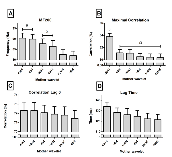
One-way repeated measures analysis of variance (ANOVARM) showed an increased peak power at 70% (221 ± 84 W) and 50% load conditions (217 ± 94 W) when compared with the 30% regime (185 ± 78 W). The one-way ANOVARM of the vastus lateralis RMS revealed no differences between load conditions (30%= 57 ± 17%, 50%= 61 ± 11% and 70%= 61 ± 14%; with p=.232). The same was found for biceps femoris RMS (30%= 33 ± 17%, 50%= 31 ± 14% and 70%= 31 ± 13%; with p=.622). The two-way ANOVARM revealed no interaction between the two main effects (Mother wavelet and Load) in any of the studied variables, regarding vastus lateralis muscle. Results of the main effect Mother Wavelet are shown in Fig. (2).
In Correlation Lag 0, Maximal Correlation and Lag Time the ANOVARM revealed significant differences (p<.05) between loads. Correlation Lag 0 showed increasing values with increasing load, being the values of the three loads statistically different (30%= 67 ± 5%, 50%= 73 ± 3% and 70%= 75 ± 4%; with p<.001). The two higher loads showed increased values of Maximal Correlation, when compared with the lower load (30%= 79 ± 4%, 50%= 82 ± 3% and 70%= 82 ± 3%; with p= p<.001). On the other side, increasing load reduced the Lag (30%= 144 ± 36 ms, 50%= 127 ± 21 ms and 70%= 108 ± 25 ms; with p=.002;). MF200 variable did not change with different loads (30%= 58 ± 17 Hz, 50%= 63 ± 14 Hz and 70%= 69 ± 23 Hz; with p=0.06).
4. DISCUSSION
The present study’s objective was to compare different mother wavelets to analyze EMG data of a power task. The main findings were: the 44th order Daubechie mother wavelet presented the highest similarity between EMG frequency and power output; there is no difference between all mother wavelets to estimate the electromechanical delay by the cross-correlation lag; and the task load did not affect the mother wavelets’ output regarding the similarity with the power output.
EMG frequency parameters were obtained by six mother wavelets in order to compare its outputs and try to pinpoint the most feasible. Significant differences were seen in both main effects, but not in the interaction. This suggests that mother wavelet selection should not be related with load condition, even though there are influence of load in EMG signal [27] and differences in mother wavelets outputs [26], as showed in our results.
In mother wavelet main effect, median frequency in the interval of 200 millisecond before peak power (MF200) and maximal cross correlation showed significant differences. Correlation at zero lag, as expected, did not showed significant differences between mother wavelets. The electromechanical delay shifts the power output curve from the EMG curve [28], losing their similarity independently of the selected mother wavelet. While the correlation at zero lag suffer a negative influence of the electromechanical delay, the lag at the maximal cross correlation value has been suggested as a good approach to identify the electromechanical delay magnitude [29]. There are no differences between mother wavelets for this parameter, meaning that the estimated electromechanical delay is independent of a particular mother wavelet function.
44th order Daubechie mother wavelet presented the highest maximal cross correlation value which means it shows the highest similarity between EMG frequency and power output curves. An optimal similarity between these two signals allows better understanding of the biological phenomena assessed. Therefore, to assess EMG time-frequency parameters in highly demand tasks in elderly mother wavelet 44th order Daubechie should be chosen.
It is interesting to notice that db44 did not show the highest MF200 value, suggesting that the curves’ similarity (power output and EMG frequency curves) does not rely on maximal or minimal discrete values. On the contrary, it seems to have a frequency content optimal value that allows obtaining a similarity between the power output and EMG frequency curves. Rafiee et al. [26] tried to find the most similar function for electromyographic, electroencephalographic and vaginal pulse signals among 324 potential mother wavelets. The authors identified db44 as the most similar mother wavelet for these classes of biosignals. The surface EMG data was obtained by 16 electrodes in the forearm of six subjects performing 10 hand movements for five seconds each: forearm pronation, forearm supination, wrist flexion, wrist extension, wrist abduction, wrist adduction, key grip, chuck grip, hand open, and a rest state. It is reasonable to assume these moments are low demanding and therefore activate mainly slow twitch fibers. In this case, their results could not fit high demanding tasks with explosive executions such as those in the present study. Nonetheless, our findings reiterates db44 as the most suitable function for analyzing frequency content in elderly’s EMG signal, in this particular task.
Regarding the second ANOVARM’s main effect, higher Maximal Correlation values were achieved with increasing loads. With higher loads the knee extension task was performed slower which allows to increase motor unit recruitment, according to Henneman's size principle [30]. The size principle states that smaller motor units are firstly recruited and then bigger ones, which in turn means that slow-twitch muscle fibers are recruited first and then fast-twitch. In the light of such assumption, our results are explained. Maximal Correlation between frequency and power curves was obtained by higher loads because it was the condition that took longer allowing the recruitment of more fast-twitch fibers producing the highest frequency curve peak. On the other hand, taking longer to extend the knees reduces the electromechanical delay, which is consistent with lower lag values. Correlation at zero lag higher values at higher loads seems to be a methodological manifestation related with the diminished lag and augmented maximal correlations in that load condition than a physiological.
It was seen that different load conditions did not change MF200. Most likely, this is more related to a methodological limitation than to a physiological phenomenon. One should realize that the median value of the frequency curve was calculated 200 milliseconds before peak power output and the frequency and power curves were shifted (lag) by 144 ms, 127 ms and 108 ms for 30%, 50% and 70% load conditions, respectively. On the other hand, selecting a window wider than 200 ms could cover the total duration of the power tasks, due to the rapid nature of the movement as the ones used in this study.
This study showed that increasing loads were followed by increasing knee extensors power output in elderly women. However, the agonist EMG amplitude signal did not present a matching trend. EMG magnitude in response to different load conditions is unaltered which in this particular test seems an expected result. Although the neuromuscular demand imposed by different load conditions is undeniably different, the nature of the task (i.e., to extend the knees as quickly as possible) is the same. Thus, it would be expected to see the same muscle activation regardless of load conditions. Pousson et al. [31] even reported lower biceps brachii RMS values in high angular velocities (240°/s) when comparing with lower velocities (60°/s) in older adults, which could be related to the time required for muscle activation. Notwithstanding, the results obtained by Klass et al. [32] are in agreement with those obtained in the present study. They also found no activation differences in ankle dorsiflexors in all tested velocities (5°/s to 100°/s). In the same way, biceps femoris RMS values were found to be alike among all load conditions, which allows applying the same rationale. Even though it is an antagonist muscle, biceps femoris activation on a knee extensor task was expected to be present. In order to increase knee stability both quadriceps and hamstrings muscles are activated [14]. This statement is even more accurate when it comes to sedentary people, like the participants in this study. Once again, the same RMS values amongst load conditions can be related to the specific nature of the task. Moreover, the lack of difference allows to assume that the previously cross correlation results were not affected by a co-contraction of the biceps femoris [14].
It seems reasonable to assume that higher load regimes increases activation of fast twitch muscle fibers and should be recognized as an optimal training load. To the best of our knowledge, this is the first study relating knee extensors activation in response to load manipulation in elderly subjects. Klass [33] suggests that the cause for decreased torque development in the elderly muscle is due to slowing of motor units contractile properties. The author refers that elderly adults may achieve tetanic fusion at lower discharge frequencies compared with young adults. Therefore, in order to recruit fast twitch muscle fibers one needs to regulate the training loads in an optimal way, which in this experimental design means with 70% of 1RM. We were unable to compare these results with current literature due to lack information about the topic.
CONCLUSION
This study constitutes a novel approach on understanding how elderly people develop knee peak power by looking upon the electromyographical signal of the major agonist muscle. Using different functions to obtain EMG time-frequency parameters yields different results and the 44th order Daubechie mother wavelet was pinpointed as the most suitable. We have also seen that different load conditions do not seems to have an influence on mother wavelet selection. Finally, higher loads yields higher knee extensors power output which does not seems to be followed by an increased vastus lateralis median frequency. Nevertheless, it seems to be an optimal training load to elderly women.
ETHICS APPROVAL AND CONSENT TO PARTICIPATE
Ethical Approval was given by the University of Sao Paulo Ethics Committee (protocol number - 2010/16).
HUMAN AND ANIMAL RIGHTS
No animals were used for this study. All humans research procedures performed in the current study were in accordance with the ethical standards of the institutional and/or national research committee and with the 1964 Helsinki declaration and its later amendments or comparable ethical standards.
CONSENT FOR PUBLICATION
All participants gave their written consent to participate in the study.
CONFLICT OF INTEREST
The author confirms that this article content has no conflict of interest.
ACKNOWLEDGEMENTS
Declared none.